Knowledge
From Microscope to Machine Vision: How to Select a Camera, Zoom Lens and Magnification
A large share of machine vision inquiries at POMEAS start the same way: a customer has been inspecting parts under a traditional microscope — 100X, 200X, 400X — and now wants to replace it with an industrial camera + zoom lens + monitor + software, ideally mounted on automated equipment. The problem is that the number they have ("400X") does not describe what a camera system needs. This guide is the conversion manual: what microscope magnification really measured, what to ask for instead, and how to select between manual zoom, motorized zoom and autofocus once the requirement is written in machine vision terms.
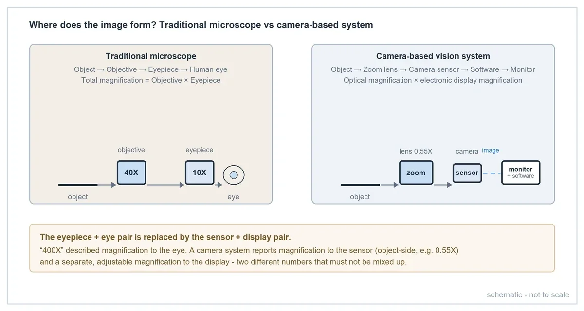
A 400X microscope does not mean a 400X machine vision lens
Traditional microscope magnification is a multiplication of two stages:
Total microscope magnification = Objective magnification × Eyepiece magnification
A "400X microscope" is typically a 40X objective looking through a 10X eyepiece: 40 × 10 = 400. That 400X describes how large the image to your eye appears compared with the object at normal viewing distance.
A machine vision system has no eyepiece. The lens forms an image directly on the camera sensor, and the "eyepiece + eye" pair is replaced by the "sensor + display" pair. So the honest answer to "I use a 400X microscope — what magnification lens do I need?" is: neither 400X nor any single number. The camera system is specified by field of view (FOV), sensor size, and the smallest feature that must be resolved. Magnification then falls out of the calculation — usually a small number like 0.55X, not 400X.
The three magnifications in a camera-based system
Because the image now passes through a display, three distinct magnifications exist. Mixing them up is the single most common source of wrong lens selection in microscope-to-camera conversions.
1. Optical magnification (object-side)
The magnification from the object to the image formed on the sensor. Formally β = image size ÷ object size. In machine vision practice it is more convenient to write:
Object-side magnification ≈ Sensor size ÷ FOV
This is the only magnification a lens datasheet can guarantee, and the only one that contributes real optical detail. It is fixed by the lens optics at a given zoom position.
2. Electronic magnification
Enlargement applied after capture: software zoom, digital crop-and-scale, pixel replication. Electronic magnification makes the image bigger on screen but adds no new optical detail — it interpolates or enlarges pixels the sensor has already captured. If a feature spans too few pixels on the sensor, no amount of software zoom will recover it.
3. Display magnification
How large the object appears on the monitor — the number users intuitively compare with "400X". Roughly:
Display magnification ≈ Optical magnification × (display width ÷ sensor width) × software zoom factor
As an illustrative geometry calculation (not a product claim): a 0.55X lens on a 2/3″ sensor shown full-screen on a monitor with a 530 mm active display width gives 0.55 × (530 ÷ 8.8) ≈ 33X apparent magnification. But a bigger monitor or deeper software zoom only changes how large the existing pixels are shown — it never increases the optical detail captured. For the zoom-vs-magnification wording itself, see zoom vs magnification: what is the difference.
Worked example: converting "400X" into a lens specification
Here is the conversion that turns a microscope habit into a machine vision requirement.
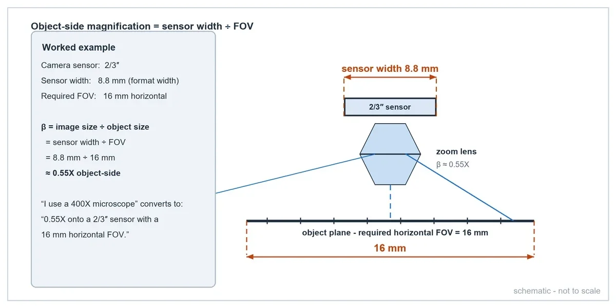
Given requirements:
- Camera sensor: 2/3″
- Required horizontal FOV: 16 mm
- 2/3″ format width: 8.8 mm (the value POMEAS uses in its FOV tables; a specific sensor's active pixel-array width can differ slightly — always divide by the actual active width from the camera datasheet)
Calculation: object-side magnification = 8.8 ÷ 16 ≈ 0.55X.
So the customer who said "I use a 400X microscope" has, once the real requirement is on the table, asked for a lens working at roughly 0.55X on a 2/3″ camera with a 16 mm horizontal field. That is a machine vision selection criterion a lens datasheet can actually answer. The step-by-step version of this conversion, with more worked math, is in Microscope 400X vs machine vision magnification: how do you convert it?
0.55X does not guarantee the microscope is replaced
This is the most important caveat on this page. The 0.55X result only answers "what magnification produces a 16 mm FOV?" It does not answer "can the system resolve the smallest feature previously visible under the microscope?" A microscope objective achieves high magnification together with high numerical aperture; a zoom lens covering a 16 mm field trades that away. Verify the whole resolution chain:
- Pixel size and camera resolution — how many pixels land on the smallest feature?
- Lens resolution — the lens must resolve finer detail than the sensor samples.
- Numerical aperture (NA) — higher NA means finer resolvable detail, shallower depth of field.
- Contrast and lighting — resolution on paper is worthless if illumination cannot make the feature visible.
- Smallest feature / defect size — the actual pass/fail criterion.
If, after this check, the smallest feature is below what a zoom lens at 0.55X can deliver, the correct answer may be a different FOV target, a higher-NA arrangement, or keeping a microscope objective in the optical path — see the section below on when a zoom lens cannot replace a microscope.
Object-side sampling: what µm/pixel really tells you
Once FOV and camera resolution are known, the object-side sampling follows immediately:
16 mm ÷ 2448 pixels ≈ 6.54 µm/pixel (for a typical 5 MP, 2448-pixel-wide 2/3-class sensor)
Each pixel corresponds to about 6.5 µm on the object. But — the second-most common conversion mistake — 6.5 µm/pixel does not mean the system reliably detects 6.5 µm defects. Reliable detection needs several pixels across the feature, optical resolution consistent with that sampling, adequate contrast, appropriate lighting and stable imaging. The sampling number is a starting point for the resolution check, not a pass/fail limit; see object-side resolution vs measurement accuracy.
When do you need a zoom lens?
A fixed-magnification FA lens is cheaper and optically stronger — if one magnification truly covers the task, use it. A zoom lens earns its place when the inspection needs more than one imaging scale on the same camera:
- Different FOVs for different products or SKU sizes
- Large-area positioning followed by close-up detail review
- Operator-adjustable observation scale at a manual workstation
- Recipe-switched magnification on an automated line
Selection then hinges on the FOV range: widest FOV, finest FOV, and the ratio between them. The mechanics are covered in the motorized zoom lens selection guide.
When do you need a motorized zoom lens?
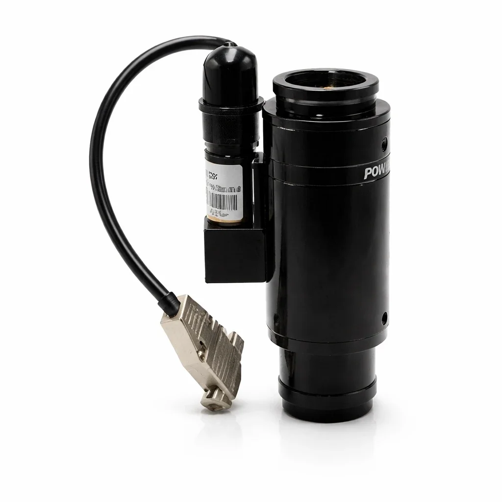
Manual zoom is fine at a bench. Once any of the following is true, the zoom axis should be motorized:
- The lens is mounted on automated equipment (robot, gantry, inline machine)
- A PLC or PC software must change FOV as part of the program
- Multiple SKUs share one station and need recipe recall
- The magnification must switch remotely or during unattended operation
Motorized models like the POMEAS LZ-650104 drive the zoom group with a closed-loop motor over RS-232, so a controller can command positions and read back status like any other machine axis. For the AI-vision flavor of this — adaptive FOV, optical recipes — see programmable optics for AI vision.
6.5X or 12.5X? Select by FOV range, not by the label
Zoom lens labels such as "6.5X" and "12.5X" name the zoom ratio — how many times the magnification range spans — not the maximum magnification. The POMEAS 6.5X LZ-650104 actually works from 0.7X to 4.5X; the 12.5X models work from 0.58X to 7.5X. What matters for selection is the FOV span those numbers produce on your sensor:
| Model | Optical mag. | Max sensor | WD | Horizontal FOV on 2/3″ (8.8 mm) |
|---|---|---|---|---|
| LZ-650104 (6.5X, motorized) | 0.7X–4.5X | 2/3″ | 82 ± 2 mm | ≈ 12.6 mm at 0.7X → 1.96 mm at 4.5X |
| PMS-LZL-12104-D1 (12.5X, motorized) | 0.58X–7.5X | 2/3″ | 77.4 ± 2 mm | ≈ 15.2 mm at 0.58X → 1.17 mm at 7.5X |
The arithmetic to apply:
- Max FOV 16 mm → min FOV 4 mm: ratio 16 ÷ 4 = 4:1 — well inside a 6.5X zoom
- 16 mm → 2 mm: 8:1 — needs the 12.5X class
- 16 mm → 1.3 mm: ≈ 12.3:1 — at the edge of the 12.5X class
And the honest check the arithmetic hides: a 16 mm horizontal FOV needs 0.55X on a 2/3″ sensor — slightly below the 0.58X wide end of the 12.5X models, so the widest achievable field is ≈ 15.2 mm. Either accept the 15.2 mm field, use a lens with a wider magnification range, or move to a larger-format sensor and lens combination. Never confirm a model on the ratio alone — verify both FOV ends against the lens's real magnification range, WD and sensor format, then confirm with the manufacturer.
When is autofocus needed?
Focus requirements follow the mechanical reality of the object plane:
- Fixed object plane, fixed WD — focus once at installation; no autofocus needed.
- Different SKUs, each at a fixed height — zoom plus stored focus presets per recipe is enough; on closed-loop motorized lenses the focus axis can be part of the same recipe recall (see parfocality and centering adjustment).
- Part height varies between captures — autofocus, driven by the vision software's contrast loop on the motorized focus axis.
- Continuous height change or moving targets — real-time autofocus must be evaluated explicitly (loop rate, search range, lens focusing travel) before promising it.
For workstations that remain microscope-like — an operator, a stand, a camera — POMEAS also offers integrated camera-based instruments such as the automatic autofocus measuring microscope, which packages the camera + zoom + autofocus conversion in one product.
When a zoom lens cannot replace a microscope objective
Some tasks genuinely need microscope optics, and saying so early saves everyone a wasted integration. A zoom lens body cannot substitute when the task needs extreme NA or sub-micron optical resolution: high-magnification biological work, oil-immersion objectives, fluorescence microscopy and other specialized techniques live in territory where illumination and contrast physics, not the magnification number, set the limit.
The datasheets make the gap concrete. The POMEAS 20X Plan APO L objective reaches NA 0.34 and 0.8 µm resolution (29.5 mm working distance, infinity-corrected). The 12.5X zoom body tops out at NA 0.110 and 3.05 µm resolution — an order of magnitude coarser. Between the two sits a practical middle path: a zoom lens used as the body with an APO microscope objective mounted in front — exactly how many industrial "digital microscopes" are built when sub-3 µm detail matters. POMEAS supplies the APO objective series for this configuration; compare zoom body and objective options or ask engineering which combination fits the feature size.
What information should you provide when replacing a microscope?
The fastest way to a correct lens selection is a complete requirement. Bring as much of this checklist as possible:
Existing microscope
- Current objective magnification and eyepiece magnification
- Current total magnification
- Current FOV, if known
Inspection object
- Object size, smallest feature, smallest defect
- Surface type (reflective, matte, curved)
Required FOV
- Maximum FOV and minimum FOV
- Whether the requirement is horizontal, vertical, or diagonal
Camera
- Camera model, sensor size, resolution, pixel size
- Mount type (C-mount, etc.)
Mechanical requirements
- Working distance, available installation height, available lens diameter
- Object height variation
Inspection requirement
- Visual observation only / defect inspection / dimension measurement / OCR / positioning / AI inspection
Automation
- Manual zoom or motorized zoom; PLC or PC control
- Preset positions, autofocus, real-time autofocus
From "I use a 400X microscope" to a selection-ready requirement
The same sentence, converted into the fields a lens engineer needs:
| Field | Converted value |
|---|---|
| Camera sensor | 2/3″ |
| Max FOV | 16 mm horizontal |
| Min FOV | [DATA REQUIRED] |
| Smallest feature / defect | [DATA REQUIRED] |
| Working distance | [DATA REQUIRED] |
| Inspection type | [DATA REQUIRED] |
| Automation | [DATA REQUIRED] |
With the first two rows filled, object-side magnification is already known (≈ 0.55X). The remaining rows decide whether a 6.5X or 12.5X zoom, autofocus, or a zoom-plus-objective arrangement is the right hardware.
Frequently asked questions
Does a 400X microscope require a 400X camera lens?
No. Microscope magnification describes the image formed for the eye (objective × eyepiece); a camera system needs the FOV, sensor size and smallest feature instead. A "400X" requirement typically converts to about 0.55X object-side on a 2/3″ sensor with a 16 mm FOV.
How do I convert microscope magnification to camera magnification?
Indirectly. Convert the microscope usage into a required FOV first, then compute object-side magnification = sensor width ÷ FOV. There is no direct formula from "400X" to a lens magnification without knowing the FOV that 400X delivered.
What magnification is needed for a 16 mm FOV?
On a 2/3″ sensor (8.8 mm format width), 8.8 ÷ 16 ≈ 0.55X. On a 1/2″ sensor (6.4 mm), it would be 0.4X. The sensor size changes the answer — which is why the camera must be specified first.
How does sensor size affect lens magnification?
Object-side magnification is sensor size divided by FOV. A larger sensor needs proportionally more magnification for the same FOV, and the lens must also cover the larger format — both the magnification and the max sensor size in the lens datasheet have to match.
Is electronic magnification the same as optical magnification?
No. Optical magnification forms a real, detail-bearing image on the sensor. Electronic magnification (software zoom) only enlarges the captured pixels, adding no new detail. Display magnification combines both.
Can a motorized zoom lens replace a microscope?
For many industrial inspection tasks, yes: camera + motorized zoom + monitor + software replaces visual microscope observation and adds automation, recipes and image storage. It cannot replace microscopy that depends on very high NA or sub-micron resolution — those tasks still need microscope objectives, alone or combined with a zoom body.
Replacing a microscope with a camera-based vision system?
Send us your current setup and we will convert it into a lens selection:
- Current microscope magnification
- Camera model or sensor size
- Maximum FOV and minimum FOV
- Smallest feature or defect to detect
- Working distance
- Manual or automated inspection requirement
POMEAS can evaluate the required optical magnification, zoom range and imaging configuration — and confirm the resolution chain before you commit to hardware.
Related reading

Subscribe to Our Newsletter
Simply enter your email to receive the latest news and insights from Pomeas. Stay connected with Pomeas and be the first to discover new innovations in optical excellence.